MP2085 - Hepatocellular Carcinoma

![]()
![]()
By buying you get
152 Points
More than a purchase. You get service and expert advice. Ask which products and combinations are recommended for you.
Clinical History
A 60-year old male is admitted with jaundice, melena and abdominal distension. He has a past medical history of untreated Hepatitis C infection from previous intravenous drug use. Further questioning reveals a 9-month history of significant fatigue, weight loss, nausea and intermittent dull right upper quadrant pain. Liver ultrasound demonstrated two large lesions within the liver. Soon after admission the patient dies from a suspected oesophageal variceal haemorrhage.
Pathology
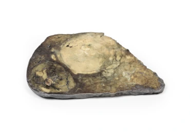
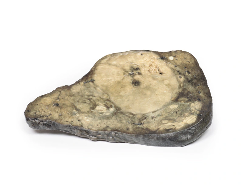
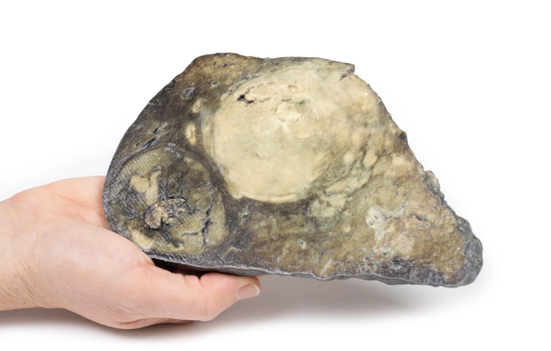
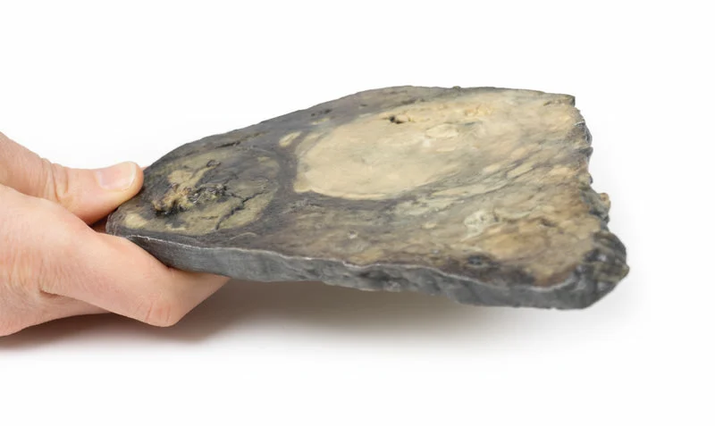
This is the liver specimen of the patient on postmortem examination. The cut surface of the liver has a multinodular appearance consistent with macronodular cirrhosis. These multiple nodules are of varying size up to 2cm in diameter, and are separated by narrow bands of fibrous tissue. There are two large round tumours also visible. These are 8cm and 6cm in diameter with a variegated cut surface due to focal necrosis, haemorrhage and bile staining. This is an example of hepatocellular carcinoma that has developed on the background of a cirrhotic liver.
Further Information
Hepatocellular carcinoma is the most common primary malignant liver cancer. HCC arises from hepatocytes in the liver. Risk factors for developing HCC include viral infections (Hepatitis B and Hepatitis C), liver cirrhosis, aflatoxin exposure, Non Alcoholic Fatty Liver Disease (NAFLD), haemochromatosis and Wilson‘s Disease. The latter is an inherited disorder in which excessive amounts of copper accumulate in the body, particularly in the liver, brain, and eyes. HCC incidence is highest in Asia and sub Saharan Africa. There is a higher risk of developing HCC in males. HCC is associated with acquired driver mutation in oncogenes and tumour suppressor genes. The two most common driver mutations that can lead to HCC are gain-of-function mutations in beta-catenin and loss-of-function mutations in p53.
Clinically HCC can present with abdominal pain, fatigue, weight loss, abdominal fullness and less commonly jaundice, gastrointestinal or variceal bleeding. HCC metastatic spread is haematogenous with lung, abdominal lymph nodes and bones being the most common extrahepatic sites. Death usually occurs from cachexia, haemorrhage or liver failure. Treatment varies on the stage of the tumour and the patient’s underlying general status and co-morbidities. Treatment can include surgical resection of ablation of the tumour, chemotherapy and liver transplantation can be curative.
- Quantitative unit
- ks

MP2085 - Hepatocellular Carcinoma