MP1755 - Cubital fossa - muscles, large nerves and the brachial artery

![]()
![]()
MP1755 - Cubital fossa - muscles, large nerves and the brachial artery
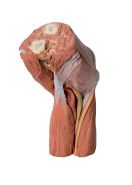
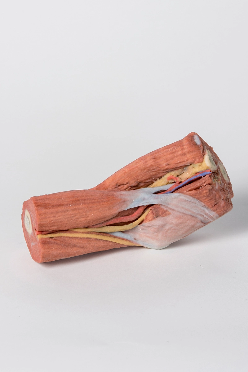
This 3D print shows the distal arm, a patially flexed elbow region and the proximal forearm. Its main purpose is to display the arrangement of muscles, large nerves and the brachial artery at the cubital fossa. All fat and superficial cutaneous nerves and veins have been removed.
Viewed from the the anterior aspect the most obvious feaure is the biceps muscle with its insertion in the form of the flattened bicipital aponeurosis passing medially over the muscles of the common flexor origin and the more rounded tendon passing deep to insert into the radial tuberosity. The brachialis muscle lies deep to biceps and is particularly obvious when looked at from the lateral aspect. In the proximal part of the forearm the brachioradialis muscle (slightly elevated and reflected laterally to reveal deeper structures) and extensor carpi radialis longus are identifiable. On the medial side one can see the classic arrangement of the biceps tendon, brachial artery and median nerve (TAN) from lateral to medial. They are partially covered by the bicipital aponeurosis as they course distally. The ulnar nerve can be seen changing position from the anterior compartment of the arm to the posterior compartment (the intermuscular septum has not been preserved but triceps muscle is clearly evident) to pass behind the medial epicondyle and enter the cubital tunnel. It travels distally between the two heads of flexor carpi ulnaris. Close inspection of the groove between brachilais and brachioradialis reveals the radial nerve which would not be visible if the brachioradialis muscle had not been partly reflected. It lies amongst some fat (yellow) but its superficial branch passes distally below brachioradialis.
Viewed from behind the most obvious structure is the triceps tendon inserting into the olecrenon process of the ulna. The medial and lateral epicondyles are also clearly visible (grey/white in colouration). The medial one is clearly identifiable as it has the ulnar nerve passing posteriorly before penetrating the deep fascia covering the gap between the two heads of flexor carpi ulnaris.
The cut surface of the arm reveals in transverse section the biceps muscle lying anteriorly with the neurovascular bundle on its medial side which contains the brachial artery together with median nerve and ulnar nerve (veins have been removed). The three heads of triceps (lateral, long and the deeper placed medial head) are clearly visible in the posterior compartment.
On the cut surface of the forearm it is more difficult to discern each muscle but the cut surfaces of the radius and ulna are clearly visible as is the brachial artery lying medial to pronator teres muscle and the median nerve lying just deep to this muscle which is the most lateral of the muscles arising from the common flexor origin.
- Quantitative unit
- ks

MP1755 - Cubital fossa - muscles, large nerves and the brachial artery