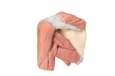
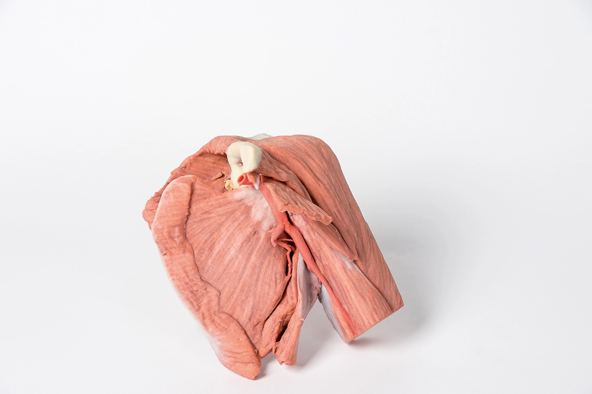
MP1523 - Shoulder (left) - Superficial muscles and axillary/brachial artery

![]()
![]()
MP1523 - Shoulder (left) - Superficial muscles and axillary/brachial artery
This printed 3D model of the left shoulder displays the superficial muscles around the shoulder joint, the rotator cuff muscles and the axillary artery as it progresses distally to become the brachial artery.
The specimen consists of the scapula, humerus and and the clavicle which has been sectioned approximately at midpoint. Inferiorly the humerus has been cut transversely at approximately at the level of the mid shaft.
The muscles attached to the clavicle have been preserved; with the subclavius muscle attachment to the inferior border of the clavicle, and the deltoid covering the lateral aspect of the proximal upper limb, concealing the origins of the long head of biceps brachii and the lateral head of triceps brachii. The clavicular head of the pectoralis major has been preserved. On the posterior aspect the superior fibers of trapezius can also be observed where they attach attached to the posterior border of the lateral third of the clavicle, and to the acromion process and the spine of the scapula. Other muscles attached to the scapula which have been preserved include the subscapularis and serratus anterior on the anterior or costal aspect. Inspection of the anterior aspect reveals that the pectoralis minor insertion onto the coracoid process of the scapula has been preserved. Posteriorly the teres major and teres minor muscles are clearly visible arising from the lateral border of the scapula. Supraspinatus is preserved but infraspinatus has partly been removed to show branches of the suprascapular artery passing from the supraspinous fossa around the base of the spine to enter the infraspinous fossa housing the infraspinatus muscle. A small stump of the attachment of omohyoid is also visible above the suprascapular ligament.
The muscles of the proximal upper limb have all been preserved, and those of the superficial layer, i.e. long head of biceps brachii, and long and lateral heads of triceps brachii, can be observed to form a complete layer of musculature around the humerus.
The axillary artery below the inferior border of the clavicle can be seen to give off the thoracoacromial branch anteriorly and just slightly more distally the suprascapular artery can be seen passing posteriorly. Coursing distally, it gives off posterior branches of the circumflex scapular and subscapular arteries. The anterior and posterior circumflex humeral arteries are hidden from view when viewed from in front, however the latter artery can just be made out deep to the posteior fibres of deltoid as it emerges though quadrangular space. Below the inferior border of teres major the axillary artery becomes the brachial artery. A radial collateral artery is visible arising from the brachial artery. The axillary artery becomes the brachial artery beyond the lower margin of the teres major muscle.
A small remnant of the suprascapular nerve passing under the suprascapular ligament is visible.
The cross section of the mid shaft of the humerus nicely displays the relations of the major neurovascular bundles and the muscles in the anterior and posterior compartments.
- Quantitative unit
- ks

MP1523 - Shoulder (left) - Superficial muscles and axillary/brachial artery